What is Antibody Feeding?
Antibody feeding is a method used to visualize receptor endocytosis and trafficking of neurotransmitter receptors.
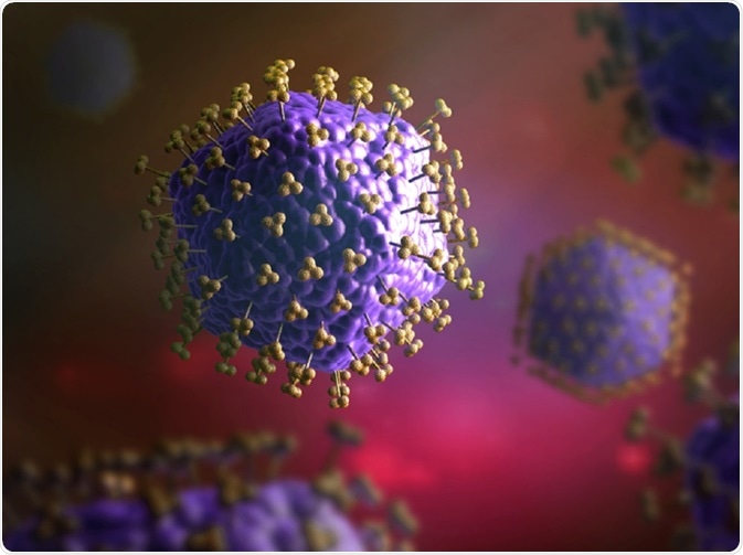
 Christoph Burgstedt | Shutterstock
How does antibody feeding work?
This method uses antibodies that can recognize and label specific extracellular epitopes on a receptor. These epitopes can be in the form of endogenous signals, such as amino acids on the extracellular domain of receptors or extracellular tags, such as myc epitope, that are engineered on the surface of recombinant receptors.
When a cell is impermeable, the antibody can bind only to surface receptors. The antibody remains bound to the receptor until the pH becomes acidic; at this time, the antibody/receptor complex dissociates and degrades. During receptor recycling, the continued bond between antibody and receptor allows for the study of receptor recycling.
Antibody binding
The binding of antibodies to cell surface receptors is performed at 4°C. Carrying out this step at this temperature blocks membrane tracking and thus, inhibits receptor endocytosis of the antibody during binding. Most cell lines can tolerate low temperatures. However, some cells, like primary cultured neurons, can be sensitive to low temperatures. Thus, some research groups perform the antibody binding assay at 37°C for this sample. In this case, the antibody is directly added to the media.
In some cases, receptors labeled with antibody can be internalized by the cell. While performing colocalization studies, blocking the uptake of receptors can be advantageous by increasing the number of receptors found on organelles. As the labelling experiments at 37°C can sometimes give rise to inaccurate results, some studies use reversible blockade of endocytosis during antibody incubation.
Endocytosis
After the antibody has bound to cell surface receptors, the cells are kept in conditioned media for endocytosis of the receptors. After receptor internalisation, cells can be kept on ice to stop endocytosis and are fixed using 2% to 4% paraformaldehyde.
The surface-receptor population can be detected by incubating cells in fluorophore-conjugated secondary antibody. The cells are then permeabilised and the receptors that are internalised can be detected using a secondary antibody that is conjugated to another fluorophore. This step can be used to separately detect surface and internalised receptors.
How are surface receptors quantified and internalized?
Advances in image processing methods and software are being used to quantify the ratio of internalised to surface receptors. This can be done by measuring and comparing the intensities of different fluorophore populations that label different receptor populations.
The imaging conditions and intensity properties should be kept constant for all imaged cells. In addition, a single fluorophore may be used to mark both populations of fluorophore along with a second fluorophore to mark only a specific population can give an internal control in the experiment.
What are the applications of antibody feeding?
Antibody feeding has been used to analyse the location of GPCRs inside the cell, and inotropic receptors and transporters on cell surfaces. The process of internalisation and trafficking of receptors can also be studied by sampling at different time points. The presence of several washing steps and harsh treatments can often make this technique less suitable for dynamic analysis.
Sources
- docksci.com/…king-of-gpcrs-new-techniq_59e648efd64ab2c25557ea05.html
- https://www.ncbi.nlm.nih.gov/books/NBK2552/
- https://bio-protocol.org/e2453
Further Reading
- All Antibodies Content
- VHH Antibodies (Nanobodies) Advantages and Limitations
- Antibody Selection using DNA Origami Scaffolds
- Uses of Histone Deacetylase (HDAC) Antibodies in Research
- Using Antibodies for Parkinson’s Disease Research
Last Updated: Jan 25, 2019

Written by
Dr. Surat P
Dr. Surat graduated with a Ph.D. in Cell Biology and Mechanobiology from the Tata Institute of Fundamental Research (Mumbai, India) in 2016. Prior to her Ph.D., Surat studied for a Bachelor of Science (B.Sc.) degree in Zoology, during which she was the recipient of anIndian Academy of SciencesSummer Fellowship to study the proteins involved in AIDs. She produces feature articles on a wide range of topics, such as medical ethics, data manipulation, pseudoscience and superstition, education, and human evolution. She is passionate about science communication and writes articles covering all areas of the life sciences.
Source: Read Full Article
