Study shows AI software improves endovascular thrombectomy treatment times for stroke patients

The implementation of artificial intelligence-powered large vessel occlusion (LVO) detection software for acute stroke triage can improve endovascular thrombectomy treatment times, according to new research from University of Texas Health Houston.
The study, which was published in JAMA Neurology, was led by co-first authors Youngran Kim, Ph.D., assistant professor of management, policy, and community health with UTHealth Houston School of Public Health; and Juan Carlos Martinez-Gutierrez, MD, a former surgery fellow in the Vivian L. Smith Department of Neurosurgery with McGovern Medical School at UTHealth Houston.
LVO occurs when a major artery in the brain is blocked. Considered one of the more severe types of strokes, an LVO accounts for an estimated 24% to 46% of acute ischemic strokes. Prompt endovascular thrombectomy—a minimally invasive surgical procedure involving the removal of a blood clot from a blocked artery in the brain—can dramatically improve outcomes in patients with LVO acute ischemic stroke; however, its efficacy is highly time-dependent.
“The benefit of endovascular thrombectomy on functional recovery is time-sensitive, so early identification of patients with strokes with large vessel occlusions, and process improvements to accelerate in-hospital care, are critical,” Kim said. “Our study has shown that the implementation of AI software has improved workflows within the comprehensive stroke centers.”
To test how to improve in-hospital endovascular therapy workflows, researchers conducted a cluster-randomized, stepped-wedge clinical trial from Jan. 1, 2021, to Feb. 27, 2022, analyzing 243 patients with LVO stroke who presented at four comprehensive stroke centers in the Greater Houston area.
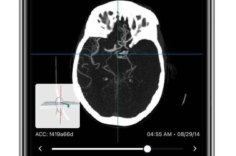
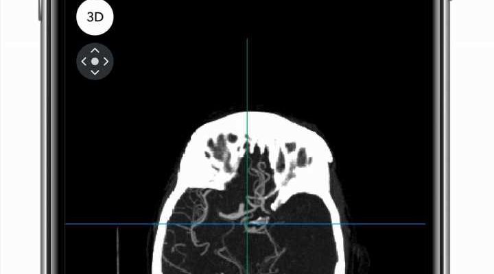
Viz LVO—artificial intelligence-enabled automated LVO detection from a computed tomography (CT) angiogram, coupled with secure messaging—was activated at the four sites in a random-stepped fashion. Once activated, clinicians and radiologists received real-time alerts to their mobile phones, notifying them of possible LVO within minutes of CT imaging completion. Members of the patient’s treatment team were also able to share information on cases in real time.
Critically, implementation of automated LVO detection software led to patients experiencing, on average, a statistically significant reduction of 11 minutes in time to thrombectomy initiation. Time from CT scan initiation to the start of endovascular therapy also fell by nearly 10 minutes.
“We are just at the beginning of automated machine-learning algorithms to benefit acute stroke care,” Giancardo said. “Many other applications are being developed, such as using CT angiograms to detect infarcted areas of the brain without advanced imaging, an NIH-funded effort by our research group which we recently published, or even the use of retina imaging as a proxy for brain imaging.”
The findings come shortly after the publication of another study led by Kim, Sheth, and other UTHealth Houston researchers, which found that, despite having worse stroke symptoms and living within comparable distances to comprehensive stroke centers, women with LVO acute ischemic stroke were less likely to be routed to the centers compared to men. Both studies are part of a broader effort to discover ways to improve stroke outcomes.
“Nearly 2 million brain cells die every minute the blockage remains, so speeding up treatments by 10 to 15 minutes can result in substantial improvements,” Sheth said. “Our study is the most rigorous of its kind to address the question of whether machine-learning software can result in a clinically meaningful improvement for patients with acute stroke, and here, we see that the answer is ‘yes.'”
More information:
Youngran Kim et al, Automated large vessel occlusion detection software and thrombectomy treatment times: A cluster randomized clinical trial, JAMA Neurology (2023). DOI: 10.1001/jamaneurol.2023.3206. jamanetwork.com/journals/jaman … jamaneurol.2023.3206
Journal information:
Archives of Neurology
Source: Read Full Article
