Exploring the effects of vascularization strategies on brain organoids

Cerebral organoids are three-dimensional, in vitro cultured brains that mimic the activities of the human brain. They have emerged as invaluable tools to comprehend evolution, disease pathogenesis, and neurodevelopmental processes.
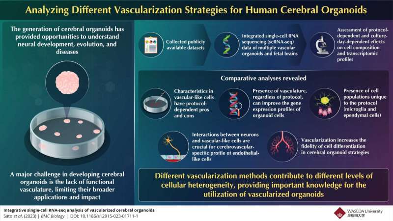
However, the development of these organoids is still in the nascent stages, with several limitations that hinder their broad applications. A major obstacle is the absence of a functional vasculature that can restrict the size of organoids, trigger cell death, and prevent cell differentiation in the organoids.
To address this, diverse strategies aiming to vascularize human cerebral organoids have been developed.
Recently, Assistant Professor Kosuke Kataoka, along with co-authors Yuya Sato and Toru Asahi from the Graduate School of Advanced Science and Engineering, Waseda University, conducted a comprehensive investigation into various strategies for vascularizing cerebral organoids.
Their study was published online in BMC Biology.
Kataoka says, “Several strategies have been proposed to construct functional vascular systems in cerebral organoids, however, no integrated comparative study of these strategies previously existed. Therefore, the characteristics and problems of each vascularization strategy have not been precisely characterized.”

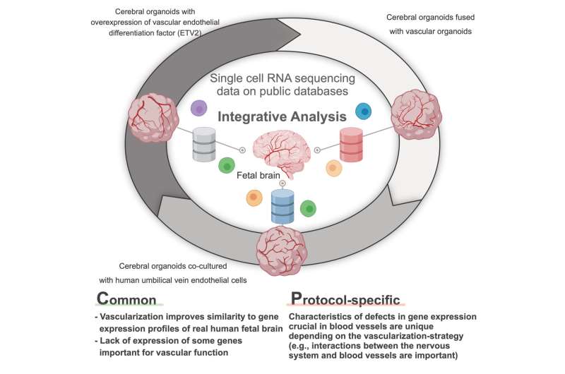
Through an integrated comparison of single-cell RNA sequencing (scRNA-seq) data, the study evaluated various vascularized human cerebral organoids created using different approaches, followed by an analysis of these datasets in conjunction with fetal brain data. This research outlined the impact of various vascularization techniques on cell type differentiation and the transcriptome profiles of both neuronal and vascular cells in these organoids.
It was observed that all the vascularization protocols improved the correlation value in most cell types. “This finding suggests that regardless of the protocol type, vascularized cerebral organoids exhibited a gene expression profile closer to the fetal human brain than non-vascularized organoids,” explains Kataoka.
They also found that vascular induction had transcriptomic effects on neuronal and vascular-like cell populations. The vascular cells of the fetal brain showed expression of all the marker genes, but the various vascularized and vascular organoids had an insufficient expression profile. Furthermore, this expression profile was found to be dependent on the vascularization strategy.
The study also revealed the importance of interactions between vascular-like cells and neurons for blood vessels to develop their cerebrovascular-specific profile to perform their vasculature functions characteristic of the brain, like the blood-brain barrier.
Expanding on the future applications of these findings, Kataoka says, “Our findings could contribute to providing more realistic human brain models with blood vessels. This will not only help in developing a better understanding of the human brain but also in accelerating the research on various brain diseases and enable more accurate drug screening.”
Vascularized cerebral organoids are unlikely to undergo cell death and, thus are thought to become the standard for future brain research.
The present research is crucial for the fabrication of vascularized organoids in the future. Here’s hoping for the development of vascularized organoids with higher fidelity.
More information:
Yuya Sato et al, Integrative single-cell RNA-seq analysis of vascularized cerebral organoids, BMC Biology (2023). DOI: 10.1186/s12915-023-01711-1
Journal information:
BMC Biology
Source: Read Full Article
